Refractive Surgeries in AL: Boost Your Vision with Professional Care
The Duty of Advanced Diagnostic Devices in Identifying Eye Disorders
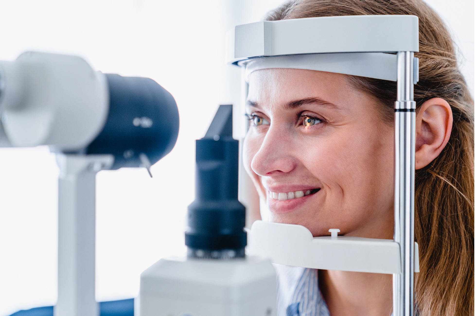
In the world of ophthalmology, the application of sophisticated analysis tools has changed the very early identification and administration of various eye conditions. As the need for exact and prompt diagnoses continues to expand, the integration of advanced tools like optical comprehensibility tomography and aesthetic field testing has become crucial in the world of eye treatment.
Value of Very Early Diagnosis
Early medical diagnosis plays a crucial role in the efficient administration and therapy of eye conditions. Prompt identification of eye problems is critical as it enables timely treatment, potentially avoiding more development of the illness and lessening long-lasting problems. By finding eye disorders at a very early phase, medical care service providers can use appropriate treatment strategies customized to the details condition, inevitably leading to better end results for individuals. Early medical diagnosis allows patients to access required assistance services and sources faster, enhancing their total quality of life.

Innovation for Finding Glaucoma
Cutting-edge analysis innovations play an important function in the early discovery and tracking of glaucoma, a leading source of irreversible loss of sight worldwide. One such innovation is optical coherence tomography (OCT), which gives in-depth cross-sectional pictures of the retina, enabling the dimension of retinal nerve fiber layer thickness. This dimension is vital in assessing damages caused by glaucoma. An additional sophisticated device is visual area testing, which maps the level of sensitivity of an individual's visual field, aiding to find any areas of vision loss attribute of glaucoma. Additionally, tonometry is utilized to measure intraocular pressure, a significant danger variable for glaucoma. This test is vital as elevated intraocular pressure can bring about optic nerve damage. In addition, newer innovations like making use of synthetic knowledge formulas in evaluating imaging information are revealing promising cause the very early discovery of glaucoma. These advanced diagnostic devices make it possible for ophthalmologists to diagnose glaucoma in its early stages, enabling prompt treatment and better management of the illness to protect against vision loss.
Function of Optical Comprehensibility Tomography

OCT's capacity to evaluate retinal nerve fiber layer density permits for precise and objective measurements, aiding in the early detection of glaucoma even before visual field problems come to be noticeable. On the whole, OCT plays a crucial role in enhancing the diagnostic accuracy and administration of glaucoma, eventually adding to much better results for individuals at danger of vision loss.
Enhancing Diagnosis With Visual Area Screening
A crucial part in comprehensive ophthalmic evaluations, visual field testing plays a critical duty in boosting the analysis procedure for various eye disorders. By assessing the full extent of a patient's visual area, this examination supplies critical info regarding the practical integrity of the entire aesthetic pathway, from the retina to the aesthetic cortex.
Aesthetic area screening is particularly valuable in the medical diagnosis and monitoring of conditions such as glaucoma, optic nerve disorders, and numerous neurological conditions that can impact vision. Through measurable measurements of outer and central vision, medical professionals can find subtle adjustments that may indicate the existence or progression of these problems, even prior to visible symptoms take place.
Additionally, aesthetic field screening permits the tracking of treatment efficiency, web link assisting eye doctors tailor healing interventions to specific patients. eyecare near me. By tracking modifications in visual area performance gradually, doctor can make educated choices about readjusting medications, suggesting medical interventions, or applying other suitable actions to protect or boost a patient's visual function
Taking Care Of Macular Deterioration

Final Thought
In conclusion, progressed diagnostic tools play an essential duty in recognizing eye disorders early on. Technologies such as Optical Coherence Tomography and aesthetic field screening have actually considerably improved the precision and efficiency of diagnosing conditions like glaucoma and macular degeneration.